The back has been hurting for a week, bending is difficult, and people around you say, “Why not get an MRI? Then you’ll know what’s going on.” In the general practitioner’s office, disappointment often follows – no referral for the scan, but rather a conversation, examination, and the advice to stay active. This is not due to cost-cutting measures. Why doctors are cautious about imaging for fresh back pain, when an MRI can actually help, and how to interpret a report full of medical jargon can be read here.
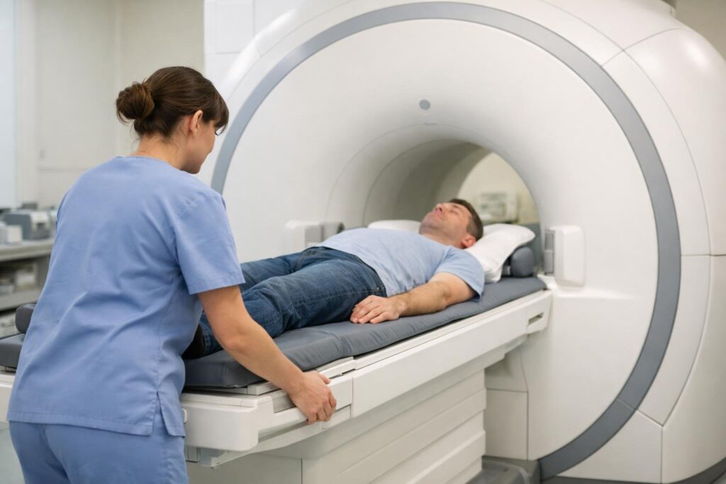
Why the General Practitioner Doesn’t Order an MRI Right Away
The National Guideline for Back Pain is unusually clear on this point: For acute and recurrent back pain, no imaging should be done as long as the conversation and physical examination do not indicate a dangerous course. This applies to X-rays and CT scans as well as MRIs.
The reason is simple. The vast majority of back pain is non-specific, meaning there is no single damaged structure that can be identified and treated on an image. The treatment remains the same, whether with or without imaging: stay active, use heat, and take pain medication for a short time if advised by a doctor or pharmacist. An image that does not change the treatment offers no benefit.
It can even be harmful.
What an MRI Shows – and What It Reveals About Pain
Magnetic resonance imaging provides a very detailed view of soft tissues: intervertebral discs, nerve roots, spinal cord, ligaments, and areas of inflammation. It uses a strong magnetic field and radio waves, without X-ray radiation. This very accuracy is the problem. The machine almost always finds something.
A review published in the American Journal of Neuroradiology in 2015 evaluated images from over 3,000 people without back complaints. Signs of disc degeneration were found in 37 percent of 20-year-olds and 96 percent of 80-year-olds. Disc bulges were present in 30 percent of 20-year-olds and 84 percent of 80-year-olds – note that these individuals had no pain. Such changes are part of the aging process of the spine, just as gray hair is part of aging in the head.
So, anyone who goes into the MRI with back pain is highly likely to receive a concerning finding. Whether this finding explains the pain is unknown. Many affected individuals become more cautious out of fear of the “damaged” disc, avoid movement, and adjust their daily lives according to the image. However, rest and fear of movement are among the factors that can turn acute pain into chronic pain. Additionally, treatments that focus on the image rather than the symptoms can be problematic.
In These Cases, Imaging is Helpful
The situation is quite different when the medical history or examination points to a specific cause. In such cases, the image answers a specific question, and the treatment depends on that answer. Imaging is particularly considered in the following situations:
- Suspicion of a pinched nerve root with paralysis or increasing weakness in the leg;
- Indications of a vertebral fracture, for example, after a fall, in known osteoporosis, or after prolonged corticosteroid treatment;
- Fever, chills, or a recent infection accompanied by severe back pain;
- A history of cancer, unexplained weight loss, or pain that worsens at night and at rest;
- Suspicion of an inflammatory rheumatic disease – typical symptoms occur before the age of 45 with pronounced morning stiffness that improves with movement.
The second reason is time. If pain continues to limit daily activities after four to six weeks despite guideline-conforming treatment or increases, the doctor should reassess whether imaging is indicated according to the guidelines. It is assessed, not automatically referred: even at this point, the examination determines whether an image can clarify an open question.
X-ray, CT, or MRI?
Which method is appropriate depends on the question. X-rays show bones – fractures, misalignments, the height of the vertebrae. Discs and nerves are not directly visible. CT scans provide a more detailed view of bones and are quickly available, but come with a higher radiation exposure. MRI is the method of choice when it comes to discs, nerves, inflammation, or tumors.
Not everyone can undergo an MRI. Certain pacemakers, implants, and metal parts in the body are obstacles or require special precautions; the radiology practice will clarify this before the appointment. If you feel anxious in tight spaces, you should mention this beforehand. The examination itself is painless but loud, and you must lie still during it.
Reading the Report Without Panic
Radiological reports describe everything that deviates from the textbook image. This can sound dramatic quickly. A few terms appear particularly often:
- Protrusion – the disc bulges forward, its outer fibrous ring is intact.
- Prolapse (herniated disc) – tissue from the soft core has protruded through the fibrous ring. This can also occur in people without complaints.
- Osteochondrosis – signs of wear on the disc and adjacent vertebrae.
- Spondyloarthritis – arthritis of the small vertebral joints.
- Spinal canal stenosis – the spinal canal is narrowed. This only becomes significant if there are corresponding symptoms, such as leg pain when walking that eases when sitting.
- “Age-appropriate” – what is common among peers. A good sign.
What is crucial is whether the image and symptoms match. If a herniation presses on the nerve root on the left, but the pain radiates into the right leg, then the herniation is an incidental finding. Therefore, have the report explained to you by the doctor who examined you. And ask directly: “Does this explain my pain? Does it change anything about the treatment?”
When to See a Doctor Immediately
With certain signs, time is of the essence. Call emergency services at 112 or seek an emergency room immediately if the following occurs alongside back pain:
- Numbness in the genital or anal area or on the inner thighs;
- Suddenly unable to hold urine or stool, or unable to empty the bladder;
- Paralysis or rapidly increasing weakness in one or both legs;
- Severe pain after an accident or fall;
- High fever and chills.
In these cases, imaging is initiated immediately – there is no waiting.
Frequently Asked Questions
Can I get an MRI at my own expense?
This is possible; some practices offer it as a self-pay service. However, this does not make the examination more meaningful. If it is medically justified, the statutory health insurance will cover the costs; if the justification is lacking, there remains the risk of an incidental finding that raises more questions than it answers.
My last MRI was two years ago. Do I need a new one?
Only if the symptoms have changed significantly. The guidelines advise against repeating images as long as the clinical picture remains the same.
The report states a herniated disc. Do I need surgery?
The image alone never decides this. The symptoms and examination findings are crucial, especially paralysis or disturbances of the bladder and bowel. Many herniations are treated without surgery; the treating doctor will discuss the options with you.
Is the doctor missing something dangerous without imaging?
Dangerous causes are rare and almost always present warning signs, which are specifically sought during the conversation and examination. If the pain does not improve as expected, a reassessment will be made.
Also Read
- Lumbago: What Helps in the First Days
- Stress Management and Burnout Prevention: Ways to Achieve Greater Well-being in Daily Life
- Prevention and Health Check-ups: Living Healthier Through Timely Measures
This article is for informational purposes only and does not replace medical advice.
Sources
- Federal Chamber of Physicians (BÄK), National Association of Statutory Health Insurance Physicians (KBV), Working Group of Scientific Medical Societies (AWMF). National Guideline for Non-specific Back Pain – Long Version, 2nd Edition. 2017. AWMF Register No. nvl-007.
- Brinjikji W, Luetmer PH, Comstock B, et al. Systematic literature review of imaging features of spinal degeneration in asymptomatic populations. AJNR American Journal of Neuroradiology. 2015;36(4):811–816.